| Parasite | Dermocystidium sp. |
|---|---|
| Taxonomy | Opisthokonta, Ichthyosporea |
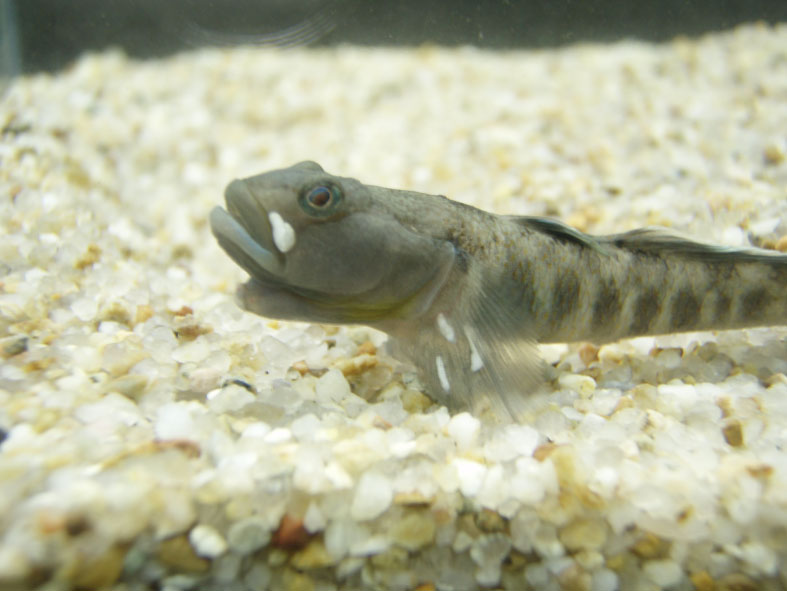
| Host | Rhinogobius sp. BW |
| Infection site | Skin, fin |
| Clinical signs | Yellowish elongated cysts are observed in the skin and fin (Fig. 1). |
| Parasitology | Previously, Dermocystidium was classified into Fungi, but now it is a protozoan (Adl et al., 2005; Hatai, 2004). A cyst is filled with many spherical spores (9.5 (9.6-12.3) mm in diameter) (Fig. 2). Strictly speaking, it is unclear whether this parasite is Dermocystidium because a spherical inclusion body and a nucleus were not confirmed in the parasite body. |
| Pathology | Unknown |
| Health hazard | Since this parasite is not infectious to human, it is harmless in food hygiene. |
| Diagnosis | Check the spherical spore by wet-mount preparation. |
| Other information | Diseased fish recovered by the cyst disruption after heating treatment. This phenomenon is similar to the case of Dermocystidium infection in Japanese eel. |
| References |
Adl, S. M., A. G. B.
Simpson, M. A. Farmer et al. (2005): The new higher level classification of
eukaryotes with emphasis on the taxonomy of protists. J. Eukaryot. Microbiol., 52 399-451. |

(Photos by Lake Biwa Museum)
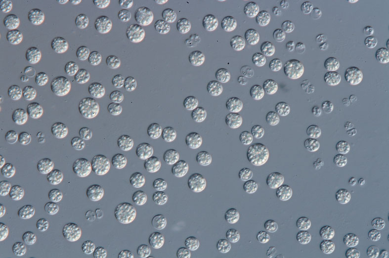
Fig. 1. Rhinogobius sp. BW infected with Dermocystidium
Fig. 2. Numerous spherical spores in the cyst.